Projects
-
Stretch Reflex Arc (Preliminary Results)
-
Description:
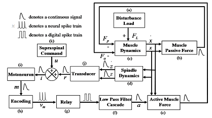
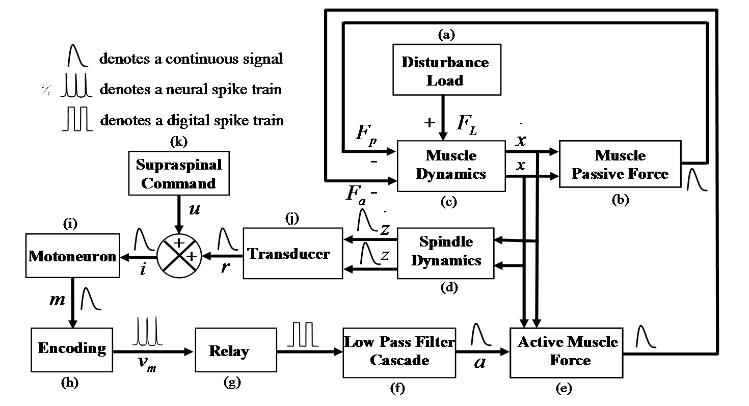
Simulink has been utilized as a tool to build and solve complex non-linear dynamic models.
A preliminary computer model of the Strech Reflex Arc has been created using in vivo
experimentally derived models for components and
subsystems from literature (Top Figure). This system is reasonably complex and consists of (i) the
motoneuron represented by a second order linear differential equation (LDE), (ii) the muscle
activation dynamics represented by a first-order LDE followed by a static saturation
non-linearity which is proceeded by a first-order non-linear differential equation (NLDE),
(iii) active and passive muscle force generation represented by static non-linear equations,
(iv) the spindle stretch dynamics represented by a first-order NLDE, (v) the encoding of
continuous mechanical motion into spike trains represented by a second order NLDE, and (v)
the DRG represented by a simple delay. The entire system is simulated as a continuous system
by a variable step mixed fourth-fifth order differential equation solver.
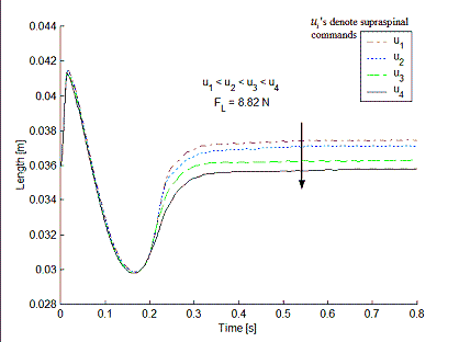
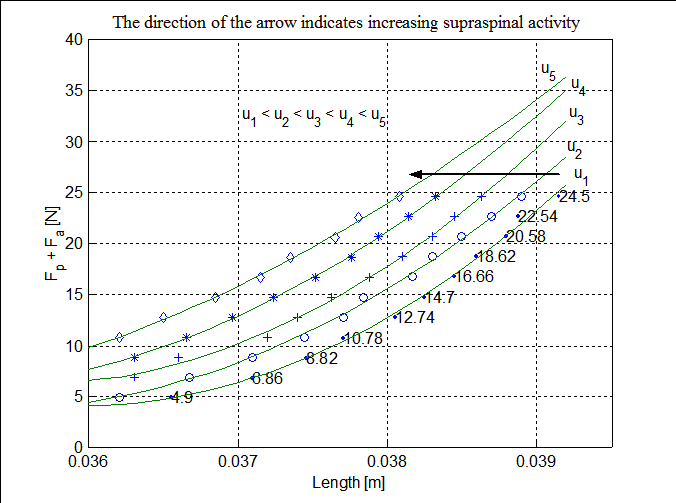
Results: Middle Figure shows the transient response of the system under a step load for
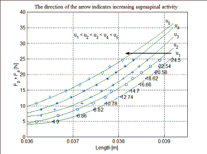
various amounts of descending control. Bottom Figure shows steady state force-displacement curves as a function
of amount of descending control. The numbers along the curve show the amount of loading for that datapoint.
-
Characterization of the Neuron Microelectrode Interface
-
Description:
A neuron encodes and expresses its response to environmental cues through a
change in the shape and/or firing rate of its action potentials. Detection and
analysis of action potential shapes and firing rates,
therefore, promises to be a mainstay of applications in high throughput functional
screening of drugs or toxins and an important indicator in ascertaining the physiological
health of long term neuronal cultures in order to monitor chronic experiments involving
neurodegenerative diseases and spinal cord injury. Conventional techniques of studying
electrophysiological activity, like intracellular patch clamping, limit the life of a
neuron to a few hours or as in the case of voltage sensitive dyes, prove to be toxic
when employed for chronic experimental studies. Another problem associated with using
voltage sensitive dyes for high throughput drug and toxin detection is that the dyes
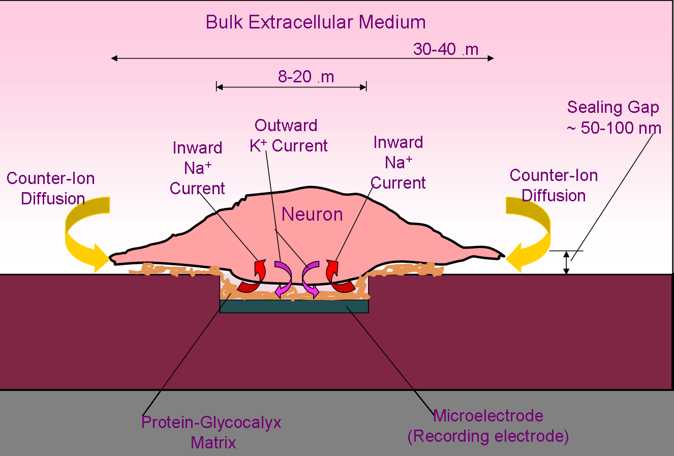
may themselves interfere with the drug or toxin chemistry. Extracellular Recordings are a viable alternative to invasive techniques but they suffer
from the following problems:
-- Low signal to noise ratio - signal attenuation due to counter-ion diffusion from the bulk extracellular medium
-- A modification of the shape of the cell-generated potentials due to the presence of a highly dispersive dielectric medium in the cell-electrode cleft.
Solution: Treat this as a nonlinear system identification problem. The following experiments
and analyses were performed:
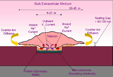
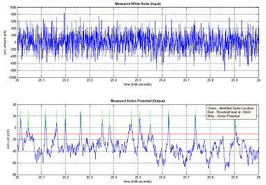
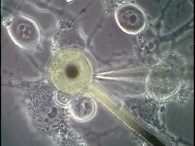
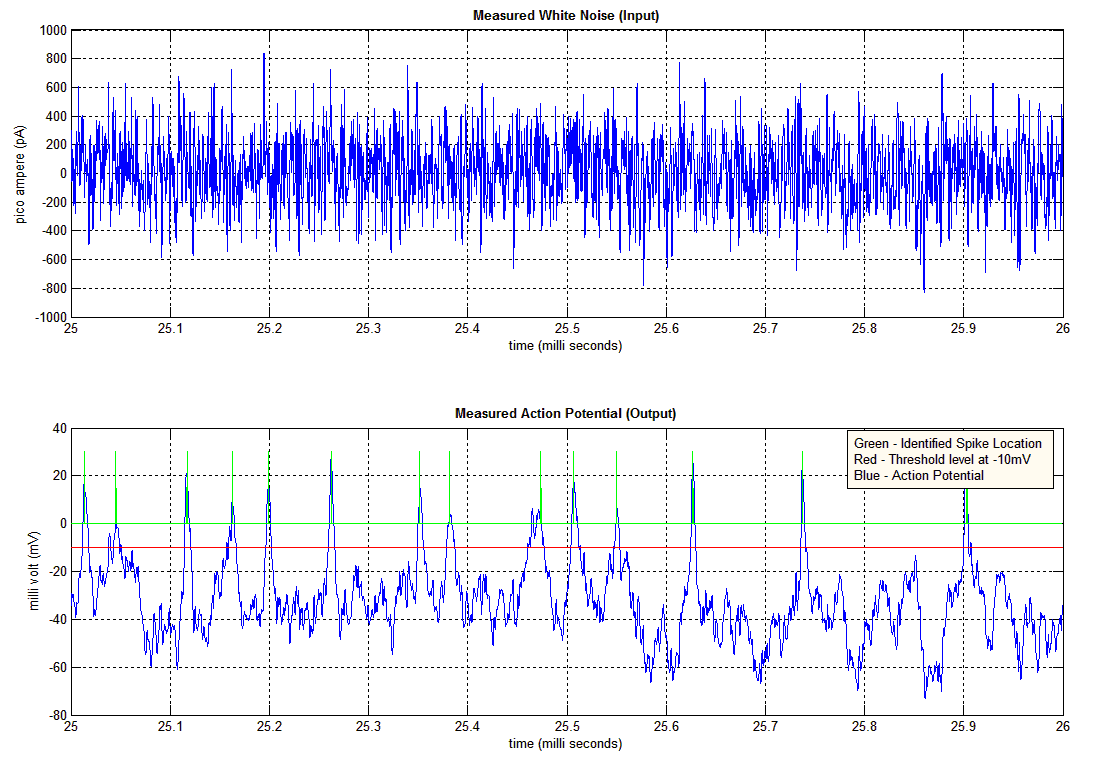
-- Stimulate system with microelectrode (See Figure: Top) under voltage clamp with white noise in amplitude and
frequency range of interest.
-- Record signal on microelectrode, appropriate signal processing to eliminate drift etc.
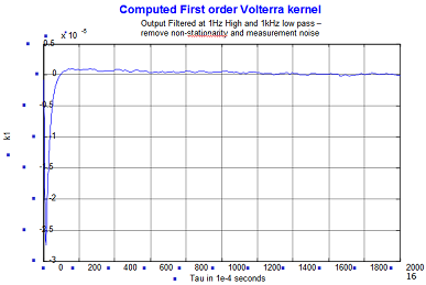
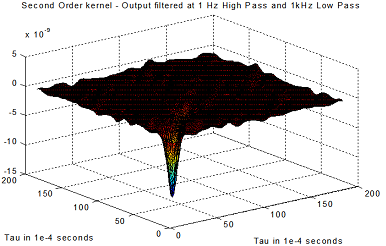
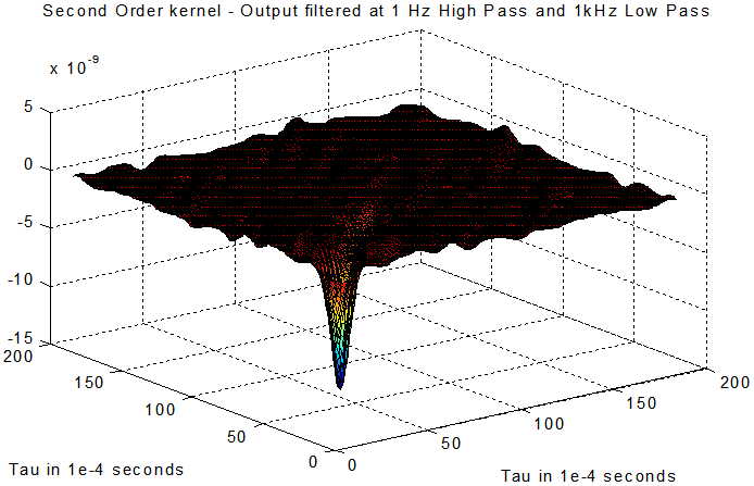
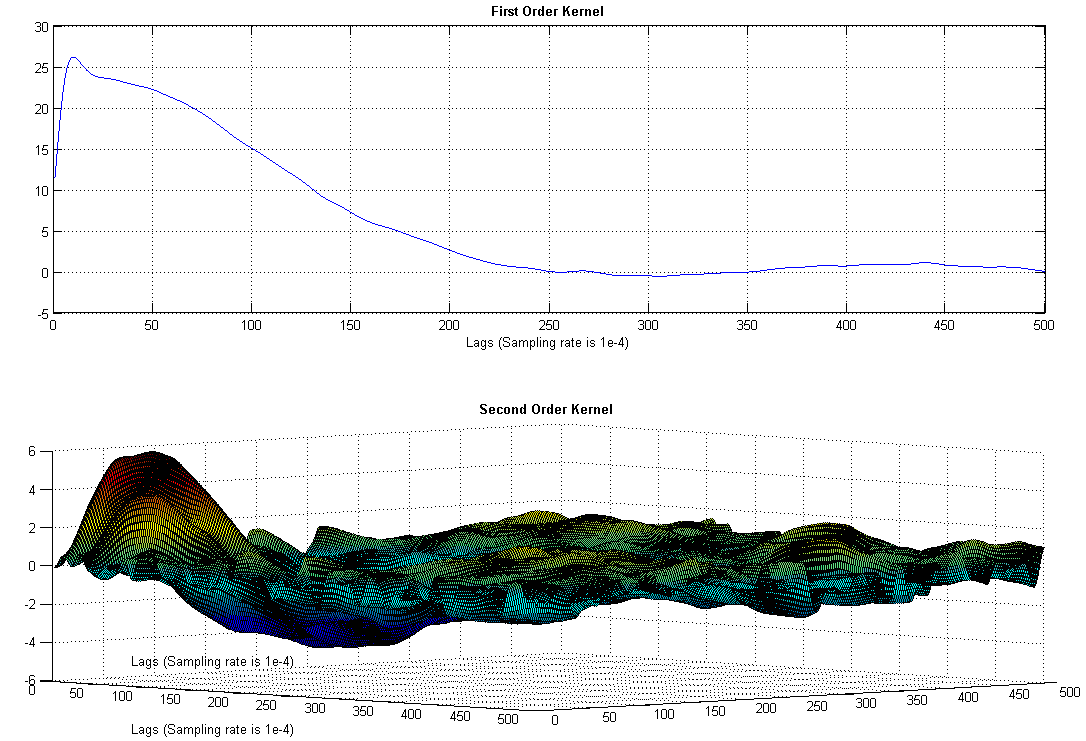
-- Compute the Volterra or Wiener kernels (See Figures for computed first and second Wiener kernels) ; to reduce
computation, discretize each kernel using Orthogonal Basis Functions (OBFs). Optimize number of OBFs for each order of
the nonlinearity.
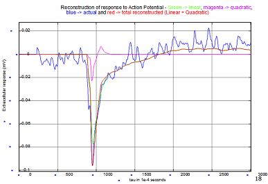
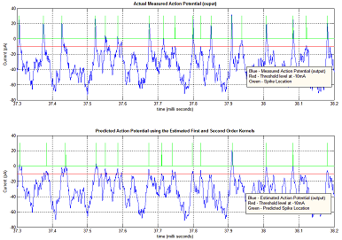
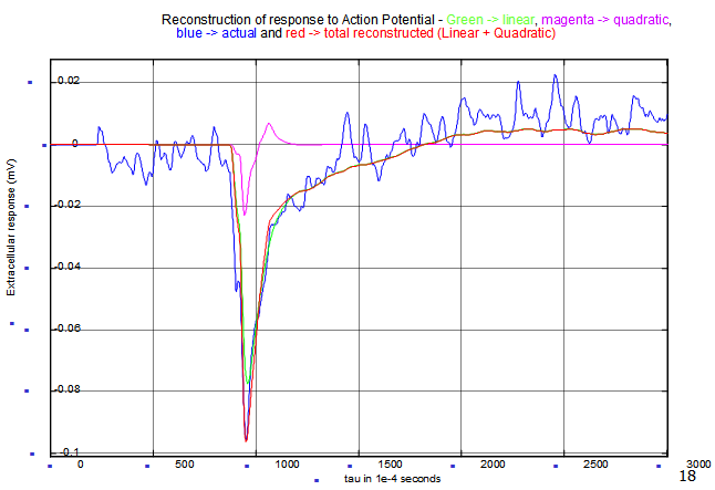
-- Stimulate system under current clamp to generate an action potential, record AP and microlectrode
signal, run the AP through the kernels and compare with experimental output to validate model. (See Figure: Bottom)
Analysis of Results:
-- Output signal is attenuated by 3 orders of magnitude, lots of counter ion diffusion.
-- System looks linear for the most part.
-- Has a nonlinearity that is substantial at higher amplitude.
-- Nonlinearity looks like inward Na and outward K currents seen during an action potential! Is it
all membrane nonlinearity or is it a combination of membrane and interface nonlinearity?
-
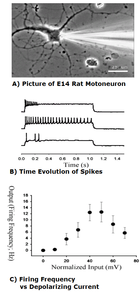
Static/Dynamic Motoneuron Modeling
|
|
|