Research

Updated: 11/04/2022

Biomedical Circuits and Systems

Our research group develops biomedical circuits and systems including electrodes, IC chips, PCB, FPGA, data acquisition systems, firmware and software! The following pictures are a few examples of the chips that we've developed for both in vitro and in vivo applications

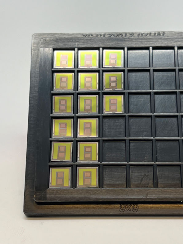
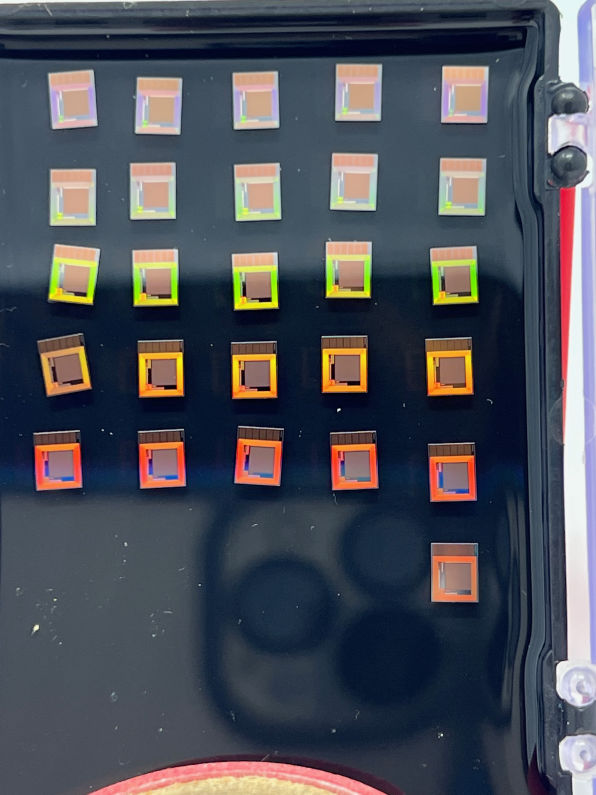
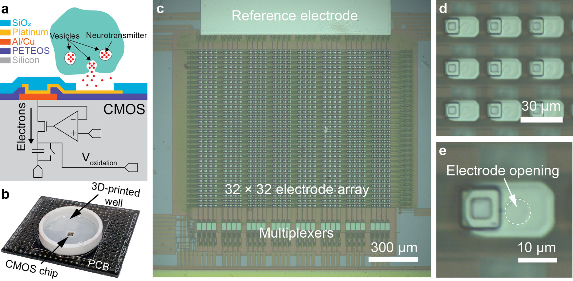
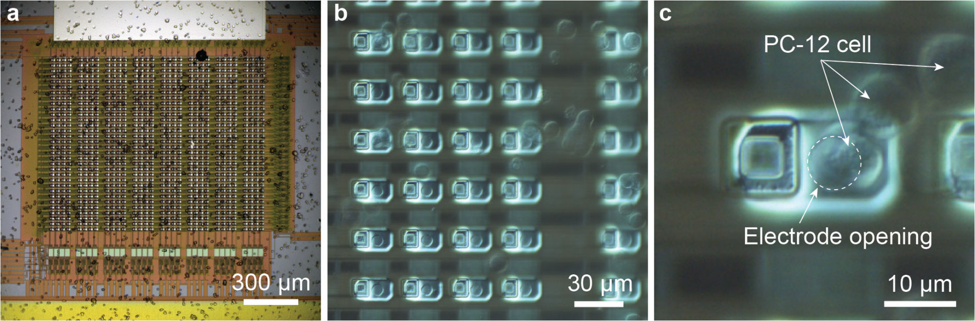
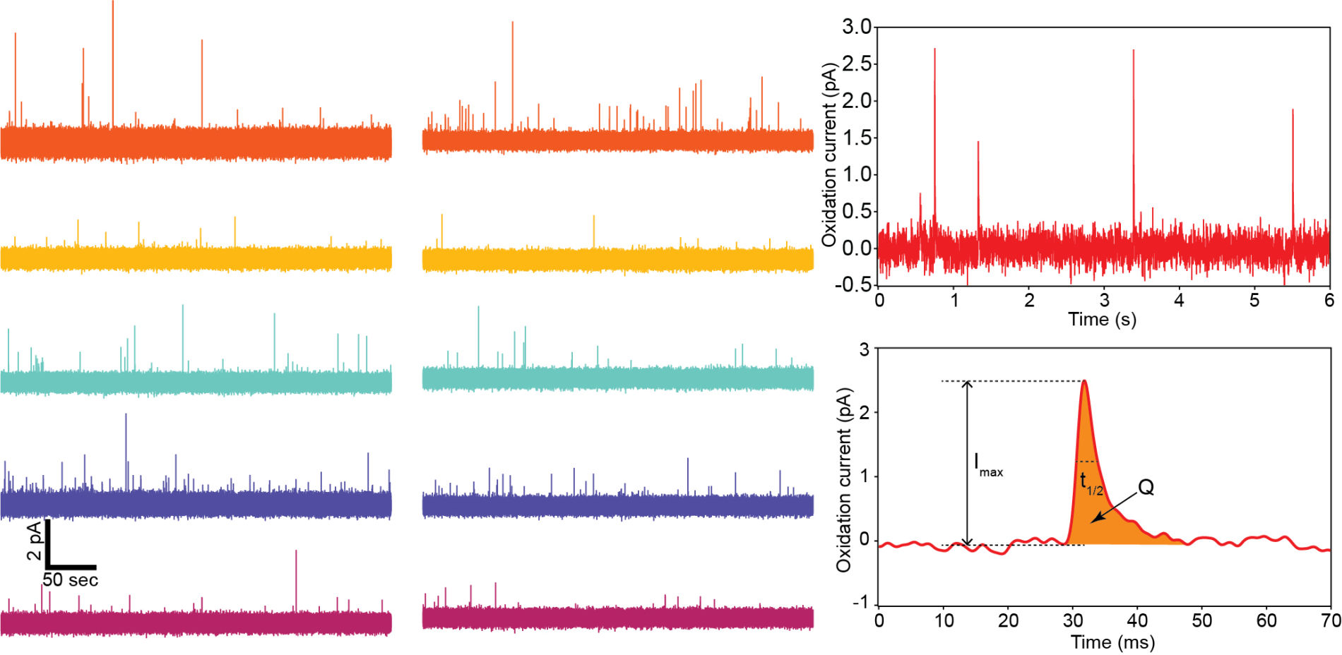
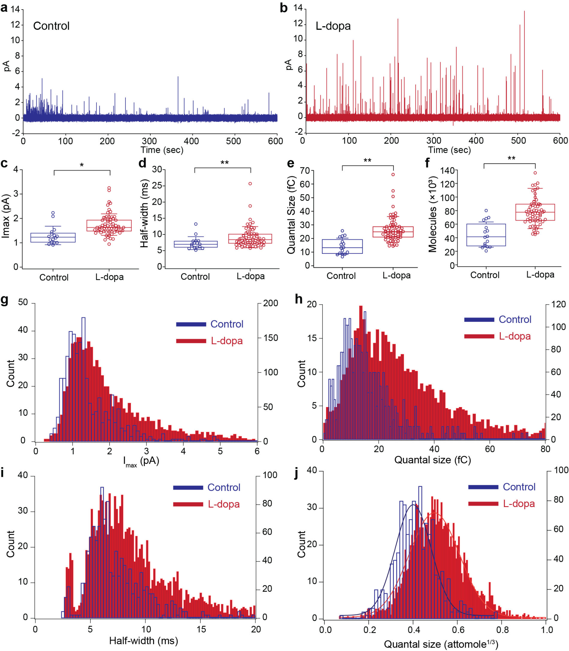
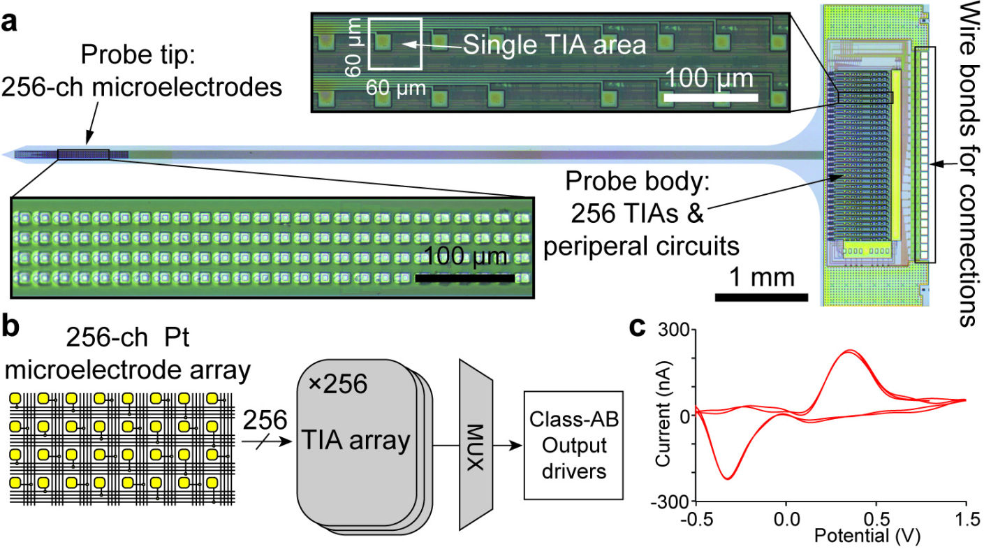
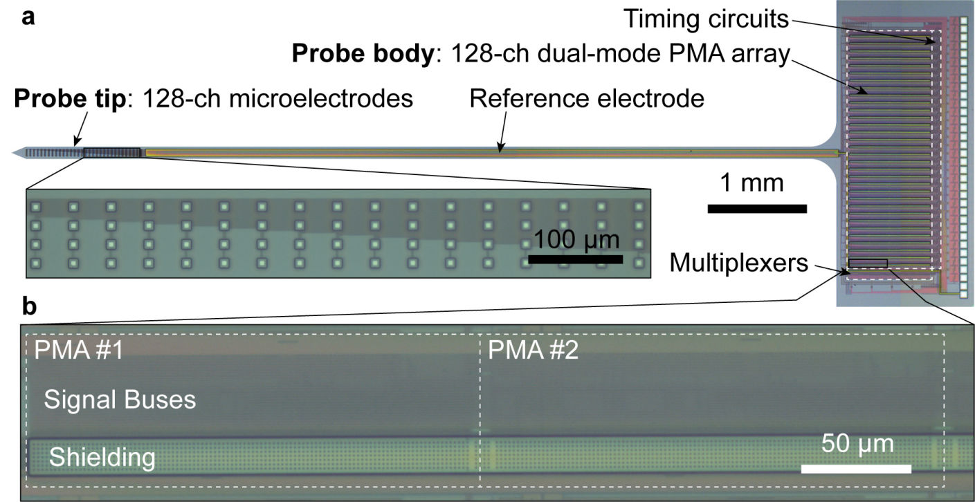
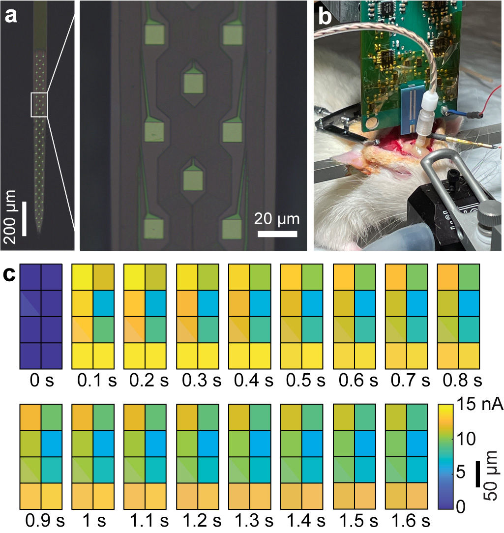
1000-ch in vitro neurochemical sensor (integrated 1024-ch on-chip electrodes + 1024 TIA amplifiers)
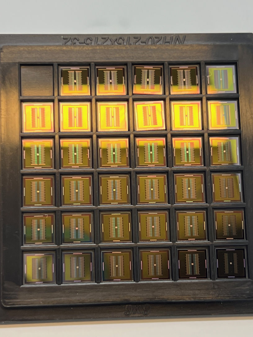
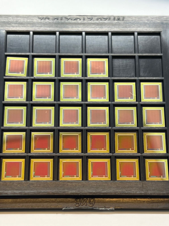
1000-ch in vitro neurochemical sensor with FSCV (integrated 1024-ch on-chip electrodes + 1024 TIA amplifiers)
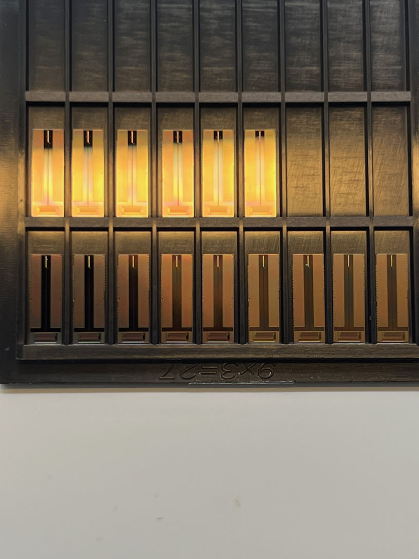
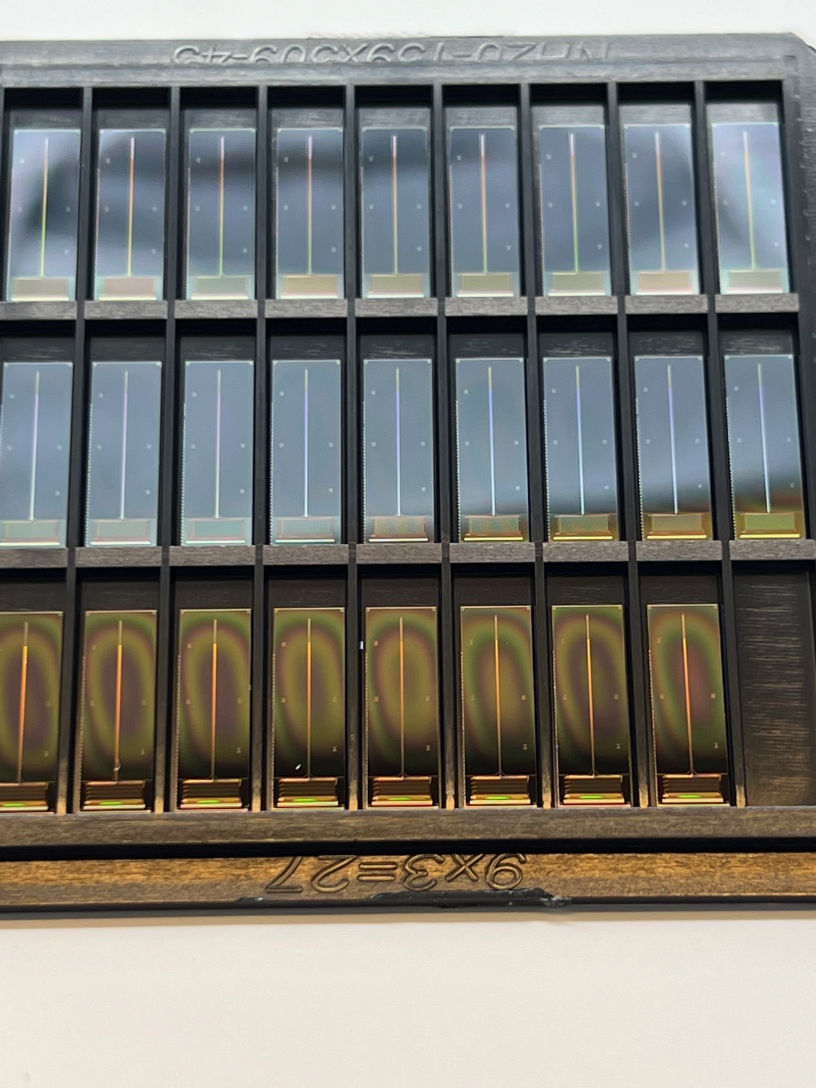
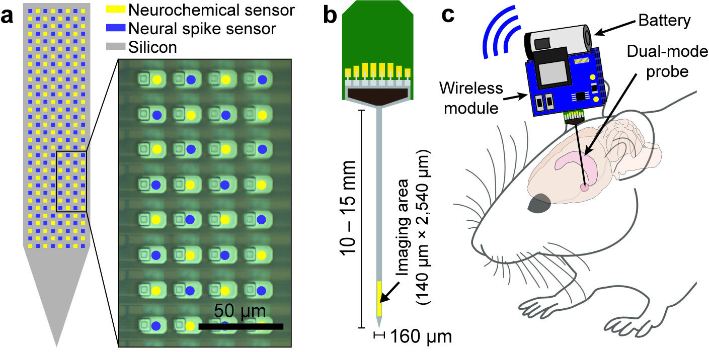
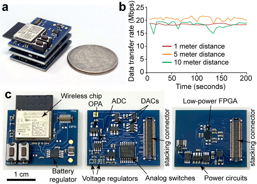
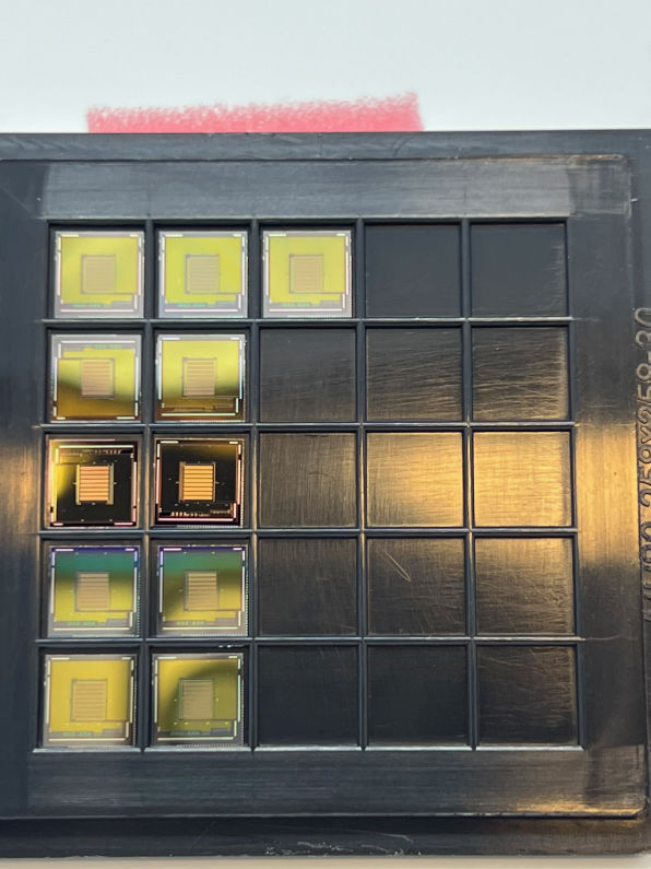
256-ch fully wireless BMI chip (integrated power coil + amplifier + electrodes)
512-ch in vitro dual-mode chip for simultaneous electrophysiology and neurochemistry (integrated 512-ch on-chip electrodes + 256 TIAs + 256 TCAs)
Fully-integrated 256-ch in vivo neurochemical probe with FSCV (integrated 256-ch on-chip electrodes + 256-ch TIA amplifiers)
Fully-integrated 128-ch in vivo dual-mode probe with FSCV (integrated 128-ch Pt on-chip electrodes + 128-ch dual-mode amplifiers)

RESEARCH AREA I: HIGH-DENSITY NEUROCHEMICAL SENSOR
NSF and DoD funded projects

Neuronal exocytosis facilitates the propagation of information through the nervous system pertaining to bodily function, memory, and emotions. Using amperometry, the submillisecond dynamics of exocytosis can be monitored and the modulation of exocytosis due to drug treatment or neurodegenerative diseases can be studied. Traditional single-cell amperometry is a powerful technique for studying the molecular mechanisms of exocytosis, but it is both costly and labor-intensive to accumulate statistically significant data. To surmount these limitations, we have developed a silicon-based electrode array with 1024 on-chip electrodes that measures oxidative signal in 0.1 millisecond intervals. Using the developed device, we are able to capture the modulation of exocytosis due to Parkinson’s disease treatment (L-Dopa), with statistical significance, within 30 total minutes of recording. The validation study proves our device’s capability to accelerate the study of many pharmaceutical treatments for various neurodegenerative disorders that affect neurotransmitter secretion to a matter of minutes.

RESEARCH AREA II: BRAIN MACHINE INTERFACE
NSF funded project

The parallel recordings from large neuron populations in the sensory cortex and primary motor cortex reveal the rich information encoded into neural signals, and guide research in restoring cognitive and motor behaviors. In such devices, the quality of information relies on the density and resolution of neural signals being measured (1). The current state-of-the-art brain-machine interface (BMI) is capable of hundreds of simultaneous electrocorticography (ECoG) recordings and was successfully used for alleviating gait deficits in primates with spinal cord injury (2–4). However, the throughput remains insufficient to be clinically relevant and significant improvements in the recording throughput are required for BMIs to help severely disabled patients to fully regain mobility or other impaired functions (5). Restoring limb movements may require a BMI to monitor 5,000 – 10,000 neurons simultaneously (6), whereas producing full-body movements may require 100,000 neural measurements (5). In recent years, many researchers are leading the BMI development to increase the throughput beyond 1000-ch recordings (2, 3, 7–10). The goal of our work is to develop a new modality that can interface with neurons.

RESEARCH AREA III: MOLECULAR DIAGNOSTICS
R01 & R21 NIH and GLA funded projects

1. 3D-manufactured real-time PCR device
Diagnosing infectious diseases using quantitative polymerase chain reaction (qPCR) offers a conclusive result in determining the infection, the strain or type of pathogen, and the level of infection. However, due to the high-cost instrumentation involved and the complexity in maintenance, it is rarely used in the field to make a quick turnaround diagnosis. In order to provide a higher level of accessibility than current qPCR devices, a set of 3D manufacturing methods is explored as a possible option to fabricate a low-cost and portable qPCR device. The key advantage of this approach is the ability to upload the digital format of the design files on the internet for wide distribution, so that people at any location can simply download and feed into their 3D printers for quick manufacturing. The material and design are carefully selected to minimize the number of custom parts that depend on advanced manufacturing processes which lower accessibility.
The presented 3D manufactured qPCR device is tested with 20-µL samples that contain various concentrations of lentivirus, the same type as HIV. A reverse-transcription step is a part of the device’s operation, which takes place prior to the qPCR step to reverse transcribe the target RNA from the lentivirus into complementary DNA (cDNA). This is immediately followed by qPCR which quantifies the target sequence molecules in the sample during the PCR amplification process. The entire process of thermal control and time-coordinated fluorescence reading is automated by closed-loop feedback and a microcontroller. The resulting device is portable and battery-operated, with a size of 12 × 7 × 6 cm3 and mass of only 214 g. By uploading and sharing the design files online, the presented low-cost qPCR device may provide easier access to a robust diagnosis protocol for various infectious diseases, such as HIV and malaria.

2. Automated, contactless, battery-operated sample preparation device
Sample preparation is an essential process that precedes nucleic acid detections which use quantitative polymerase chain reaction (qPCR). However, sample preparation is a labor-intensive process and requires skilled labor, thus limiting the public’s access in low-resource settings to many high-quality nucleic acid-based detection mechanisms. In this paper, we present a simple, handheld, battery-operated sample preparation device to minimize user’s involvement. The device uses a simple pouring method to process the DNA sample without pipetting or using disposable pipette tips. The developed device has a size of 12 × 8 × 8 cm3 and mass of only 364 g. The device is compared to gold standard methods, including magnetic bead-based and silica filter-based DNA extractions. For a short segment DNA target of 68 bp, the presented device captured 8.67× more DNA compared to that of the manual magnetic bead-based method. Because of automation, the measured capture efficiency is more consistent and has a smaller deviation between multiple repetitions than the manual method. To present a comprehensive, portable, battery-operated diagnostic system, the sample preparation device is tested in conjunction with a 3D-manufactured qPCR device. The test using three diluted target DNA samples, each spiked in whole blood (1×, 0.1×, and 0.01×), revealed a quantitative detection with ideal cycle threshold separations between the measurements. The combination of two devices will aid in resource-limited settings to promptly and accurately diagnose infections of patients.

CONTACT

Email: brian.kim at ucf.edu
Address: HEC-339/BSBS-450

AFFILIATION
  • Department of Electrical and Computer Engineering
  • College of Engineering

JOINT AFFILIATION
  • Burnett School of Biomedical Sciences
  • College of Medicine